
Gliomas are diagnosed using a neurological assessment, brain imaging, biopsy of the tissue, and molecular testing. For most gliomas, an MRI scan can give clinicians information about the location, size, and characteristics of the tumor, and investigation of tumor tissue can confirm a diagnosis and provide information about the precise kind of glioma. Today the diagnosis of a tumor is more and more a combination of the histological features seen at the microscope and the molecular genetic features.
This integrated approach is significant because two tumors with similar imaging appearances may behave differently depending on the molecular profile. Accurate classification helps clinicians to predict prognosis and determine the best treatment strategy.
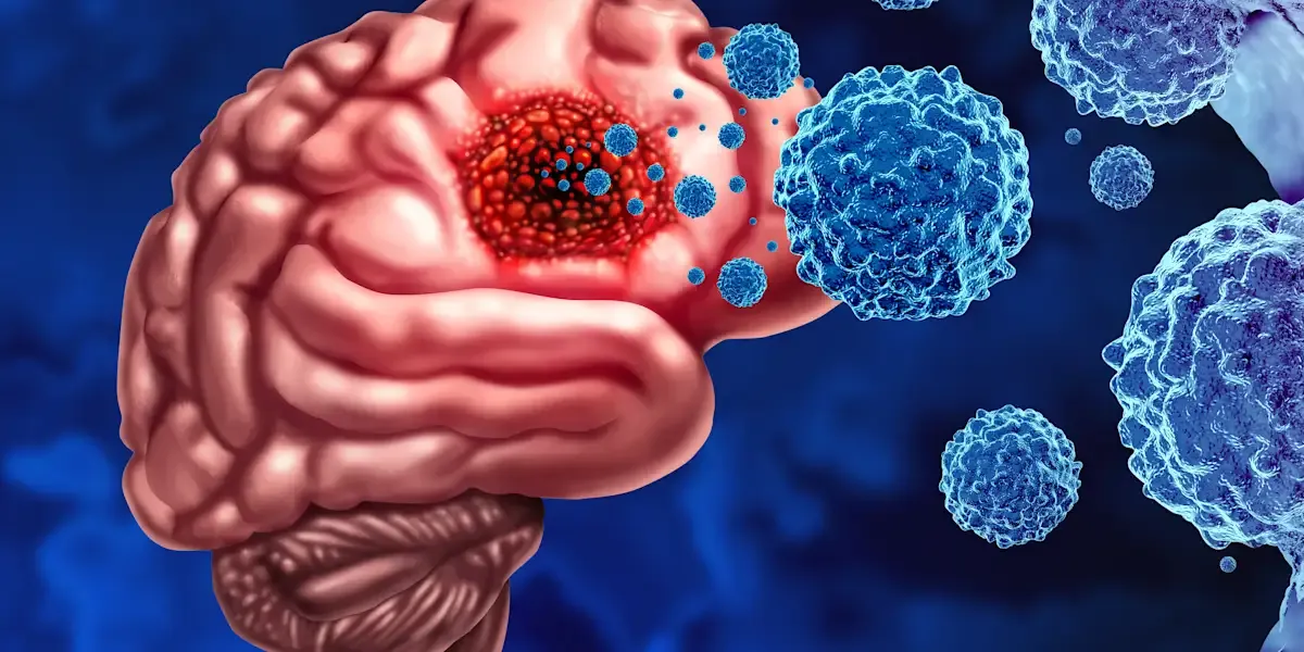
Symptoms Leading to Evaluation
The symptoms of a glioma very much depend on the location of the tumor. The warning signals may be:
• Headaches that are new or worsening
• Convulsions
• Nausea and/or vomiting
• Difficulty thinking or remembering
• Alters in speech
• Eye troubles
• Numbness or weakness
• Difficulty walking or balancing
• Behaviour or personality changes
Sometimes a seizure, especially a first unexplained seizure in an adult, might lead to imaging that reveals a brain tumor.
Medical & Physical Review
Doctors start by examining the patient’s symptoms and medical history. A neurological exam can check vision, hearing, reflexes, coordination, balance, muscle strength, sensation, memory, and mental function.
The findings can provide hints about which part of the brain might be involved, but a single examination cannot definitively diagnose a glioma.
Main Diagnostic Tests
The main tools used to diagnose gliomas are:
• Neurological examination:
• MRI
• CT scan in some cases
• Tumour Surgical Biopsy or Removal
• Histopathological
• Genetics and Molecular Testing
Instead of simply confirming that a mass is abnormal, these tests are used in combination to determine what the tumor is.
Imaging
If a brain tumor is suspected, MRI is usually the most important imaging test. The contrast-enhanced MRI can give you a lot of detail regarding the size, location, relationship to nearby structures in the brain, and other features of the tumor.
CT scans may also be used, especially in emergencies or if an MRI cannot be done. Although imaging can be highly suggestive of a glioma, it generally cannot provide the whole molecular diagnosis on its own.
Biopsy / Tissue examination
A precise diagnosis sometimes involves examination of the tumor tissue. Diagnosis may be made at the time of surgery to remove the tumor or by stereotactic biopsy when complete removal is not possible or desirable.
A neuropathologist looks at the sample under the microscope, studies the tumor cells, and characterizes them. Then we combine tissue analysis with molecular discoveries to give an integrated diagnosis.
Laboratory / Molecular Testing
Molecular testing is now a staple in modern glioma diagnosis.
Doctors may look for biomarkers such as IDH mutations, 1p/19q codeletion, and other molecular alterations that are relevant, depending on the suspected tumor. For example, the diagnosis of an oligodendroglioma needs both an IDH mutation and a 1p/19q co-deletion. MGMT promoter methylation may also have substantial implications for prognosis and therapy decisions in some gliomas.
Staging or Classification
Gliomas are not normally staged in the same way as many malignancies with a regular Stage I to Stage IV approach.
Instead, they are characterized by tumor type and CNS WHO grade and defining molecular features. This classification helps in predicting the behavior of the tumor and in planning treatment.
Further Tests
The doctor may also do other tests of speech, memory, thinking, hearing, sight, or movement depending on the location and symptoms of the tumor.
In some circumstances, advanced imaging techniques may be employed to further characterize the tumor or to aid in surgical planning.
Differential Diagnosis
Not all abnormal brain lesions are gliomas.
Doctors have to differentiate gliomas from other primary brain tumors, metastatic malignancies, infections, inflammatory illnesses, vascular abnormalities, or non-cancerous neurological problems. This is another reason why imaging alone may not be adequate to make a conclusive diagnosis.
Common Misconceptions
"An MRI alone can tell you the specific type of glioma."
Not typically, no.
MRI is important, but often tissue and molecular testing are necessary for an accurate, integrated diagnosis.
"All gliomas are diagnosed and treated the same."
Nope.
Gliomas are a heterogeneous group of tumors. Molecular characteristics, location, grade, and patient-specific factors all can have a significant impact on diagnosis and treatment.
"Gliomas are staged like any other cancer."
Not really.
Gliomas are classed according to tumor type, molecular features, and WHO CNS grade, rather than the traditional cancer staging schemes used for many other tumors.
FAQs (Frequently Asked Questions)
1. What is the first test to detect a glioma?
MRI is the principal imaging technique for the detection and characterization of a suspected glioma, although a conclusive diagnosis generally requires tissue and molecular analysis.
2. Do I need to have a biopsy?
In general, tissue diagnosis is necessary, but the mode of diagnosis depends on aspects such as tumor location, surgical accessibility, and clinical status of the patient.
3. Blood test to diagnose glioma?
To date, routine blood tests cannot be used to make a conclusive diagnosis of a glioma. Diagnosis is mostly based on imaging and tumor tissue analysis.
Must Read: How is infertility in women diagnosed?
